The homunculus
 he little man is the most reproduced picture in neuroscience: a body drawn across the surface of the brain, with huge lips, hands and tongue and a tiny trunk. It comes from the operating theatre. Between the 1920s and the 1950s Wilder Penfield operated on hundreds of people with epilepsy in Montreal under local anaesthetic, touched points on the exposed cortex with an electrode, and noted what moved or what the patient felt.
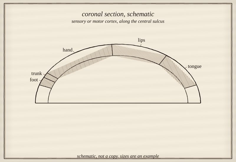
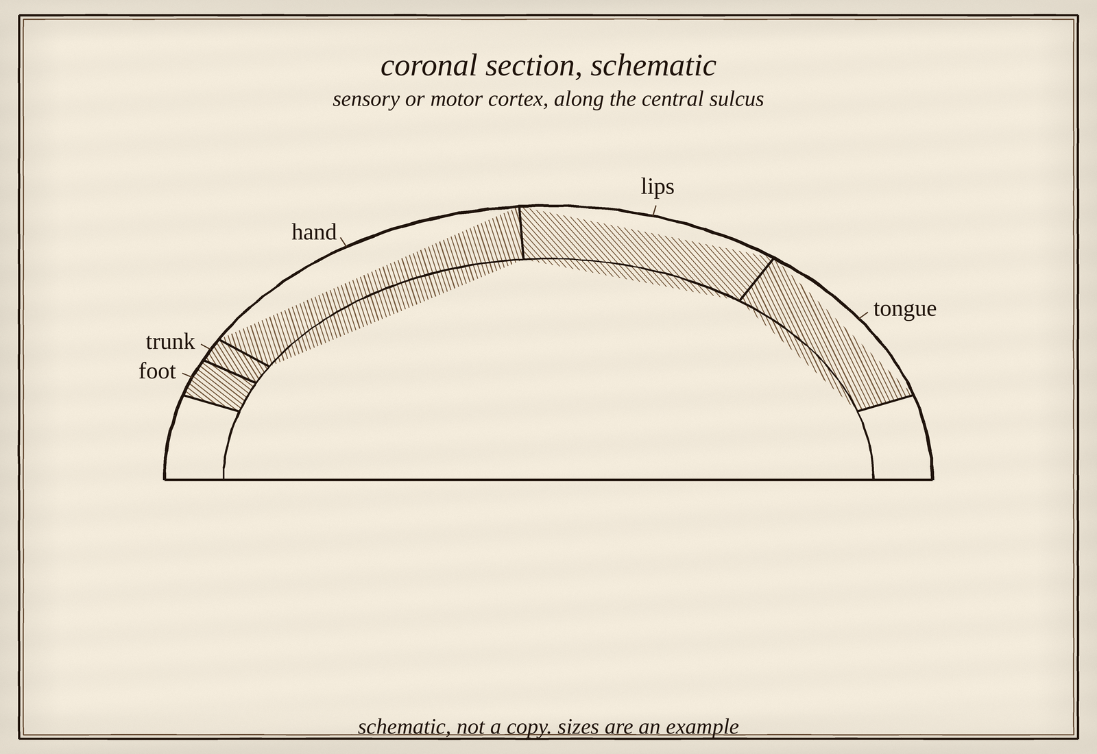
Schematic strip of sensory or motor cortex along the central sulcus, blocks for foot, trunk, hand, lips and tongue
Not a copy of Penfield's figures. Blocks, in order: foot, trunk, hand, lips, tongue. Foot, hand and mouth are the zones Gordon and colleagues (2023) name; hands, lips and tongue large, trunk and foot small. Sizes are an example. Drawn by script. credits |
how it was drawn
Penfield and Boldrey (1937) published the first summary figure, a little man whose parts are laid along the central sulcus. The famous versions, a motor and a sensory figure draped over a cross-section of the hemisphere, appeared in Penfield and Rasmussen (1950) and were drawn by a medical illustrator. Schott (1993) traces how the images were made and what liberties they take; Catani (2017) gives the longer history.
what the map hides
The drawing is a summary of many patients, and it looks more orderly than the data. Neighbouring parts overlap; individuals differ. Graziano and colleagues (2002) stimulated monkey motor cortex for half a second instead of a few milliseconds and evoked whole coordinated actions, a hand brought to the mouth, rather than twitches of one part: perhaps a map of actions rather than of body parts.
Gordon and colleagues (2023), using precise imaging in individual people, found that the strip is interrupted: separate zones for foot, hand and mouth alternate with regions belonging to a network tied to planning whole-body action, which they called the somato-cognitive action network.
The little man is still useful. It is also a reminder that the pictures neuroscience keeps are the ones that are easy to draw.
in the tour: the room called "The map" has a placard called "What the map hides" ···
Sources
- Penfield & Boldrey (1937). Somatic motor and sensory representation in the cerebral cortex of man as studied by electrical stimulation. Brain. doi:10.1093/brain/60.4.389
- Penfield, W. & Rasmussen, T. (1950). The Cerebral Cortex of Man: A Clinical Study of Localization of Function. New York: Macmillan.
- Schott (1993). Penfield's homunculus: a note on cerebral cartography. Journal of Neurology, Neurosurgery & Psychiatry. doi:10.1136/jnnp.56.4.329
- Catani (2017). A little man of some importance. Brain. doi:10.1093/brain/awx270
- Graziano, Taylor & Moore (2002). Complex Movements Evoked by Microstimulation of Precentral Cortex. Neuron. doi:10.1016/s0896-6273(02)00698-0
- Gordon et al. (2023). A somato-cognitive action network alternates with effector regions in motor cortex. Nature. doi:10.1038/s41586-023-05964-2
see also
<< all oddities